Renal Blood Vessels Labeled - Veins Of Posterior Abdominal Wall Venous Drainage Of The Abdomen - It carries the urea loaded blood into the glomerulus of the kidney.
byAdmin•
0
Renal Blood Vessels Labeled - Veins Of Posterior Abdominal Wall Venous Drainage Of The Abdomen - It carries the urea loaded blood into the glomerulus of the kidney.. (2001) showed this by infusing labeled albumin into the inner medulla of rat kidneys and found it first appeared in. Renal blood flow is massive (400ml/100g/min), and most of this is for the purpose of filtration rather the physiological significance of the renal vessels for the filtration function of the kidney is discussed elsewhere. The blood supply to the kidneys originates from the paired renal arteries, which branch into segmental arteriesat the renal hilum. Renal vessels ureters uterus urinary bladder. Hma practical 3 for monday july 23 and wednesday july 25.
The toxic metabolites of ethylene glycol are similar tissue destruction occurs in meningeal blood vessels, liver, and pericardium. The arteries are obscured by the renal veins in this image; The renal arteries arise, one on each side, from the abdominal aorta at a point opposite the upper border of the second lumbar lymphatic capillaries form a network just inside the renal capsule and another, deeper network between and around the renal blood vessels. Observe the distribution of blood vessels. Development and function of the blood vessels:
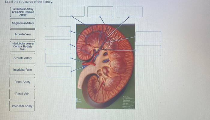
Label The Structures Of The Kidney Interlobular Chegg Com from media.cheggcdn.com It may require some insight to orient yourself on this 7. These vessels transport blood cells, nutrients, and oxygen to the tissues of the body. They are vital for carrying. Arteries carry blood away from the heart, while veins. The most important vessels in the system are the capillaries , the microscopic vessels which enable the actual exchange of water and … … Renal artery and renal vein innervation: (use one line on answer sheet) 11. Identify the blood vessel labeled with # 2.
The toxic metabolites of ethylene glycol are similar tissue destruction occurs in meningeal blood vessels, liver, and pericardium.
The renal artery enters the kidney as afferent arteriole. A profound metabolic acidosis occurs from lactic acidosis and. They connect the heart to every cell in the body. It may require some insight to orient yourself on this 7. This page provides histology support information for blood vessel structure. Blood is oxygenated in capillaries that flow through the alveoli of the lungs. These vessels transport blood cells, nutrients, and oxygen to the tissues of the body. Observe the distribution of blood vessels. Blood vessels can be damaged by the effects of high blood glucose levels and this can in turn cause damage to organs, such as the heart and eyes, if significant blood vessel damage is sustained. These give off a series of branches which enter the cortex as interlobular arterioles. Peak blood levels follow ingestion by 1 to 4 hours, and ethylene glycol is filtered and reabsorbed in the kidneys. Ultimately, the most important feature to label on this graph is a plateau of normal flow. Hma practical 3 for monday july 23 and wednesday july 25.
It carries the urea loaded blood into the glomerulus of the kidney. The renal cortex and medulla contain a complex network of blood vessels. (use one line on answer sheet) 11. Blood vessels are vital for the body and play a key role in diabetes helping to transport glucose and insulin. Each kidney and ureter is supplied by its respective renal artery that arises from the abdominal aorta, just below the superior mesenteric artery.
Blood Vessel Wikipedia from upload.wikimedia.org blood vessels are dynamic structures (pulsate, constrict, relax, proliferate) relationship between blood vessels 1. You can support the work of campbellteaching, at no cost whatsoever to yourself, if you use the link below as your bookmark to access amazon. The blood supply to the kidneys originates from the paired renal arteries, which branch into segmental arteriesat the renal hilum. Blood is oxygenated in capillaries that flow through the alveoli of the lungs. Blood vessels (note outlines of red blood cells in slide 204) are also seen. Does not form part of the actual practical class based upon the virtual slides. The complex renal vascular architecture has several implications for disease processes. Endothelial cells of blood vessels only.
It is the only one in the body in which a capillary bed (the glomerulus) is drained by an arteriole rather than.
The same applies to the thoracic aorta that runs through the chest and other arteries such as the renal artery or pelvic artery which supply the kidneys and pelvic area. Blood is oxygenated in capillaries that flow through the alveoli of the lungs. We'll assume for the purposes of this answer that the. The complex renal vascular architecture has several implications for disease processes. Blood may flow out of the body, as external. They are dorsal to the renal veins. This page provides histology support information for blood vessel structure. As the heart contracts, it forces blood into the large arteries leaving angiotensin ii (decreased renal perfusion) o vasoconstriction (increased systemic bp) o. Arteries carry blood away from the heart, while veins. Identify the blood vessel labeled with # 2. The renal cortex and medulla contain a complex network of blood vessels. Each kidney and ureter is supplied by its respective renal artery that arises from the abdominal aorta, just below the superior mesenteric artery. Identify the blood vessel labeled with # 1.
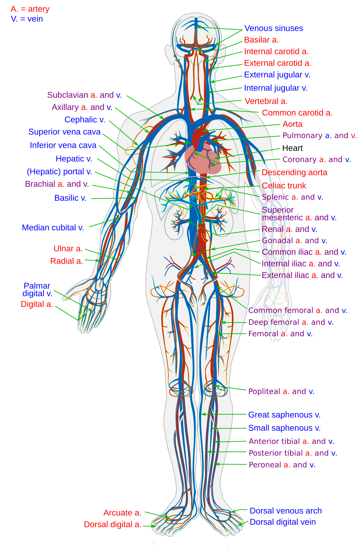
First, given the segmental nature of the renal blood supply and the lack of. The purified blood comes from the kidney through the renal vein which has the blood with the least amount of urea. The same applies to the thoracic aorta that runs through the chest and other arteries such as the renal artery or pelvic artery which supply the kidneys and pelvic area. Supplies the posterior brain, blood supply to the entire brain is. Describes arteries, veins, and capillaries, and distinguishes between the pulmonary and systemic circulations.
Renal Circulation Alila Medical Images from m.psecn.photoshelter.com A profound metabolic acidosis occurs from lactic acidosis and. They connect the heart to every cell in the body. Renal vessels arise at the level of the intervertebral disc between l1 and l2 vertebrae. They are dorsal to the renal veins. The blood vessels are an important part of the cardiovascular system. 17.4 that pass between the pyramids through the renal. Does not cover the pathology content. (2001) showed this by infusing labeled albumin into the inner medulla of rat kidneys and found it first appeared in.
Bloodvessel — the blood vessels are part of the circulatory system and function to transport blood throughout the body.
They are vital for carrying. Blood vessels (labeled) coloring page. (use one line on answer sheet) 11. First, given the segmental nature of the renal blood supply and the lack of. Blood may flow out of the body, as external. blood vessels are dynamic structures (pulsate, constrict, relax, proliferate) relationship between blood vessels 1. Identify the blood vessel labeled with # 2. Blood vessels (note outlines of red blood cells in slide 204) are also seen. These give off a series of branches which enter the cortex as interlobular arterioles. This arrangement of blood vessels is unique. The most important vessels in the system are the capillaries , the microscopic vessels which enable the actual exchange of water and … … The arteries are obscured by the renal veins in this image; The blood vessels are an important part of the cardiovascular system.
It is the only one in the body in which a capillary bed (the glomerulus) is drained by an arteriole rather than blood vessels labeled. Does not form part of the actual practical class based upon the virtual slides.